10 Oct articles, news Stem Cell Therapy Could Be Breakthrough Against Type 1 DiabetesOctober 10, 2023 By مدیرسایت 0 comments Facebook Twitter Pinterest linkedin WhatsApp Telegram WEDNESDAY, Oct. 4, 2023 -- People with type 1 diabetes lack functional islet cells in their pancreas to produce the hormone insulin a...Continue reading
18 Sep articles, news Introduction and treatment of burn woundsSeptember 18, 2022 By مدیرسایت 0 comments Facebook Twitter Pinterest linkedin WhatsApp Telegram There are many types of burn wounds caused by thermal, radiation, chemical, or electrical contact. Thermal burns. These burns ...Continue reading
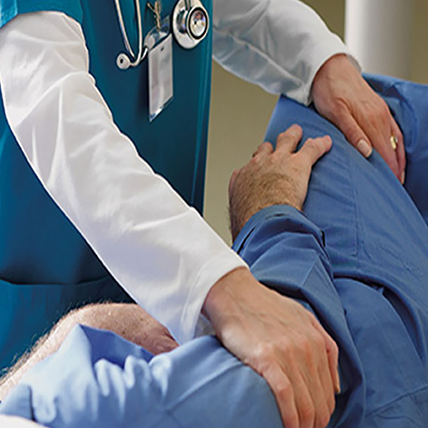
07 Sep articles, news wound vacuum therapy in traumatic woundsSeptember 7, 2022 By مدیرسایت 0 comments Facebook Twitter Pinterest linkedin WhatsApp Telegram Traumatic wounds include a group of acute, generally extensive, wounds with loss of cutaneous lining, associated or not with fracture...Continue reading
23 Aug articles, news Negative pressure therapy for the pressure ulcersAugust 23, 2022 By مدیرسایت 0 comments Facebook Twitter Pinterest linkedin WhatsApp Telegram Pressure ulcers (PU) are caused by the pressure maintained between a bone prominence and the patient's bed, leading to ischemia and n...Continue reading
14 Aug articles, news Printing technique creates effective skin equivalent, heals woundsAugust 14, 2022 By مدیرسایت 0 comments Facebook Twitter Pinterest linkedin WhatsApp Telegram Chronic wounds are deep and difficult to repair. Often, the top of the injury heals before the bottom, so the wound collapses in on i...Continue reading
03 Aug articles, news Choose pressure level for all types of wounds with Simplex III devicesAugust 3, 2022 By مدیرسایت 0 comments Facebook Twitter Pinterest linkedin WhatsApp Telegram In the following cases, a pressure more than -125 mmHg and less than -150 mmHg can be used: High secretions wounds Vacu...Continue reading
20 Jul articles, educational videos, news choose therapy mode in wound vacuum therapyJuly 20, 2022 By مدیرسایت 0 comments Facebook Twitter Pinterest linkedin WhatsApp Telegram JTNDZGl2JTIwaWQlM0QlMjI3NjYwNDY1NzMzNSUyMiUzRSUzQ3NjcmlwdCUyMHR5cGUlM0QlMjJ0ZXh0JTJGSmF2YVNjcmlwdCUyMiUyMHNyYyUzRCUyMmh0dHBzJTNBJTJGJTJ...Continue reading
17 Jul articles, news The first human trials of scarless wound healing creamJuly 17, 2022 By مدیرسایت 0 comments Facebook Twitter Pinterest linkedin WhatsApp Telegram Researchers from The University of Western Australia (UWA; Perth, Australia) have partnered with industry to conduct a world-first st...Continue reading
27 Jun articles, news Oral nutritional supplements effective for hard-to-heal woundsJune 27, 2022 By مدیرسایت 0 comments Facebook Twitter Pinterest linkedin WhatsApp Telegram Specialised oral nutritional supplement can be a therapeutic option for hard-to-heal wounds, conclude Adriano A Mehl (Universidade Te...Continue reading
03 Aug articles Creative Closure of Tunneling and Undermining Wounds with white foamAugust 3, 2021 By مدیرسایت 0 comments Facebook Twitter Pinterest linkedin WhatsApp Telegram Wounds treated with negative pressure wound therapy (NPWT) are not often straightforward. They occur in interesting places, have anythi...Continue reading