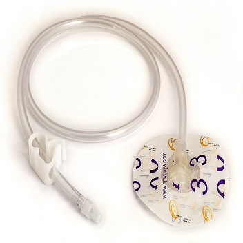
۰۹ اسفند اخبار, مقالات SPP دو لوله، سیستم پیشرفته انتقال و تخلیه همزمان در وکیومتراپی زخماسفند ۹, ۱۴۰۴ نویسنده مدیرسایت ۰ comments Facebook Twitter Pinterest linkedin WhatsApp Telegram SPP دو لوله یک ست تخصصی انتقال مایع در سیستمهای وکیومتراپی دارای قابلیت شستشو (Irrigation) است که با طراحی دو مجرای مجزا، امکان تزریق...ادامه مطلب
۲۸ بهمن اخبار, مقالات اهمیت توزیع یکنواخت فشار در وکیوم تراپی و نقش SPPبهمن ۲۸, ۱۴۰۴ نویسنده مدیرسایت ۰ comments Facebook Twitter Pinterest linkedin WhatsApp Telegram مقدمه وکیومتراپی زخم (Negative Pressure Wound Therapy) یکی از مؤثرترین روشهای نوین در انواع زخم ها محسوب میشود. این روش با ایجاد فش...ادامه مطلب
۱۲ بهمن اخبار, مقالات محفظه وکیومتراپی زخم: کنترل ایمن ترشحات، افزایش اثربخشی درمانبهمن ۲۸, ۱۴۰۴ نویسنده مدیرسایت ۰ comments Facebook Twitter Pinterest linkedin WhatsApp Telegram محفظه جمعآوری ترشحات 800 سی سی، یکی از اجزای کلیدی سیستم وکیومتراپی زخم (NPWT) است که با هدف جمعآوری، نگهداری و کنترل بهداشتی ترشحات...ادامه مطلب
۱۲ بهمن اخبار, مقالات کیت های وکیوم تراپی زخم در ۳ سایز متناسب با انواع زخمبهمن ۲۸, ۱۴۰۴ نویسنده مدیرسایت ۰ comments Facebook Twitter Pinterest linkedin WhatsApp Telegram کیت وکیوم تراپی زخم: (Negative Pressure Wound Therapy Kit) راهکار کامل، ایمن و استاندارد برای مدیریت پیشرفته زخم کیت وکیومتراپی ز...ادامه مطلب
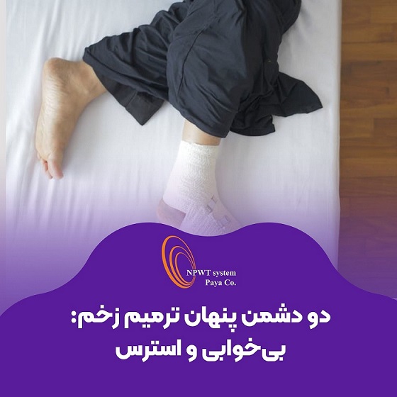
۳۰ تیر اخبار, مقالات خواب کافی و مدیریت استرس؛ نیمی از درمان زخمتیر ۳۰, ۱۴۰۴ نویسنده مدیرسایت ۰ comments Facebook Twitter Pinterest linkedin WhatsApp Telegram مقدمه ترمیم زخم یک فرایند پیچیده و چندوجهی است که تحت تأثیر عوامل مختلفی از جمله تغذیه، وضعیت ایمنی بدن، نوع زخم، سن بیمار و شرایط روا...ادامه مطلب
۲۱ تیر اخبار, مقالات زخم دیابتی چیست؟ علت، علائم، روشهای درمان و پیشگیری کاملتیر ۳۰, ۱۴۰۴ نویسنده مدیرسایت ۰ comments Facebook Twitter Pinterest linkedin WhatsApp Telegram زخم دیابتی یکی از شایعترین عوارض بیماری دیابت است که بیشتر پای بیماران را درگیر میکند. این مشکل میتواند کیفیت زندگی فرد را به شد...ادامه مطلب
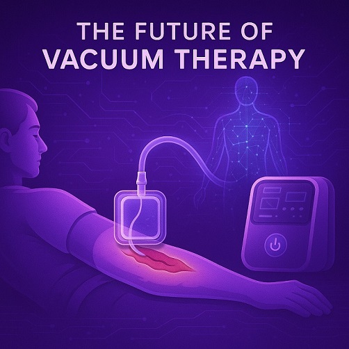
۲۵ فروردین اخبار, مقالات آینده روشن وکیوم تراپی؛ انقلابی در درمان زخمهای مزمناردیبهشت ۲, ۱۴۰۴ نویسنده مدیرسایت ۰ comments Facebook Twitter Pinterest linkedin WhatsApp Telegram مقدمه در دنیای امروز، با افزایش بیماریهای مزمن و مشکلاتی همچون دیابت، زخمهای فشاری و زخمهای جراحی، نیاز به روشهای درمانی نوین بی...ادامه مطلب
۲۲ مهر مقالات زخم بستر، بهترین روش های پیشگیری و درمانمهر ۲۲, ۱۴۰۳ نویسنده مدیرسایت ۰ comments Facebook Twitter Pinterest linkedin WhatsApp Telegram زخم بستر زخم بستر، که به آن زخم فشاری نیز گفته میشود، نوعی آسیب پوستی است که به دلیل فشار مداوم بر روی یک ناحیه مشخص از پوست ایجا...ادامه مطلب
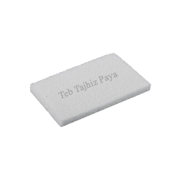
۰۴ مهر اخبار, مقالات فوم سفید، پانسمان قدرتمند در انواع زخم هامهر ۴, ۱۴۰۳ نویسنده مدیرسایت ۰ comments Facebook Twitter Pinterest linkedin WhatsApp Telegram پانسمان فوم سفید نوعی پانسمان است که از فوم PVA ساخته شده و برای پوشش و حفاظت زخمها استفاده میشود. این پانسمانها بهخصوص در درمان زخ...ادامه مطلب
۲۱ شهریور اخبار, مقالات همه چیز درباره پانسمان هیدروکلوئید + مقایسه با سایر پانسمان هاشهریور ۲۱, ۱۴۰۳ نویسنده مدیرسایت ۰ comments Facebook Twitter Pinterest linkedin WhatsApp Telegram پانسمان های هیدروکلوئیدی(Hydrocolloid dressings) یک محیط مرطوب و عایق برای ترمیم زخم فراهم می کند که برای تسریع بهبود زخمها و جلوگیری ...ادامه مطلب