۰۹ اسفند اخبار, مقالات SPP دو لوله، سیستم پیشرفته انتقال و تخلیه همزمان در وکیومتراپی زخماسفند ۹, ۱۴۰۴ نویسنده مدیرسایت ۰ comments Facebook Twitter Pinterest linkedin WhatsApp Telegram SPP دو لوله یک ست تخصصی انتقال مایع در سیستمهای وکیومتراپی دارای قابلیت شستشو (Irrigation) است که با طراحی دو مجرای مجزا، امکان تزریق...ادامه مطلب
۲۸ بهمن اخبار, مقالات اهمیت توزیع یکنواخت فشار در وکیوم تراپی و نقش SPPبهمن ۲۸, ۱۴۰۴ نویسنده مدیرسایت ۰ comments Facebook Twitter Pinterest linkedin WhatsApp Telegram مقدمه وکیومتراپی زخم (Negative Pressure Wound Therapy) یکی از مؤثرترین روشهای نوین در انواع زخم ها محسوب میشود. این روش با ایجاد فش...ادامه مطلب
۱۲ بهمن اخبار, مقالات محفظه وکیومتراپی زخم: کنترل ایمن ترشحات، افزایش اثربخشی درمانبهمن ۲۸, ۱۴۰۴ نویسنده مدیرسایت ۰ comments Facebook Twitter Pinterest linkedin WhatsApp Telegram محفظه جمعآوری ترشحات 800 سی سی، یکی از اجزای کلیدی سیستم وکیومتراپی زخم (NPWT) است که با هدف جمعآوری، نگهداری و کنترل بهداشتی ترشحات...ادامه مطلب
۱۲ بهمن اخبار, مقالات کیت های وکیوم تراپی زخم در ۳ سایز متناسب با انواع زخمبهمن ۲۸, ۱۴۰۴ نویسنده مدیرسایت ۰ comments Facebook Twitter Pinterest linkedin WhatsApp Telegram کیت وکیوم تراپی زخم: (Negative Pressure Wound Therapy Kit) راهکار کامل، ایمن و استاندارد برای مدیریت پیشرفته زخم کیت وکیومتراپی ز...ادامه مطلب
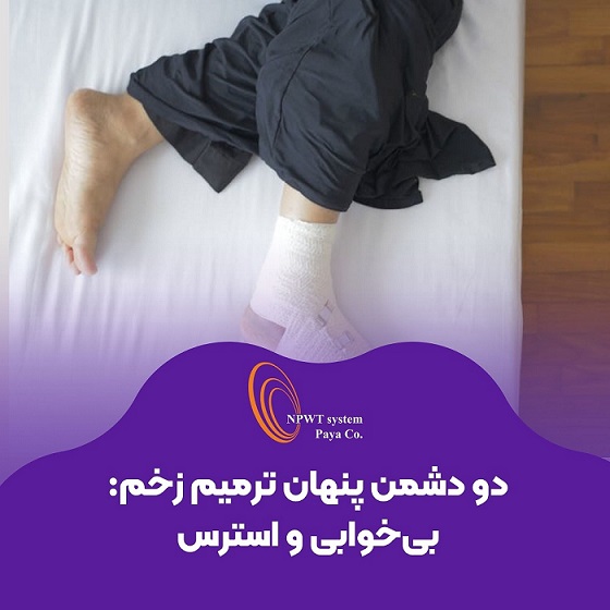
۳۰ تیر اخبار, مقالات خواب کافی و مدیریت استرس؛ نیمی از درمان زخمتیر ۳۰, ۱۴۰۴ نویسنده مدیرسایت ۰ comments Facebook Twitter Pinterest linkedin WhatsApp Telegram مقدمه ترمیم زخم یک فرایند پیچیده و چندوجهی است که تحت تأثیر عوامل مختلفی از جمله تغذیه، وضعیت ایمنی بدن، نوع زخم، سن بیمار و شرایط روا...ادامه مطلب
۲۱ تیر اخبار, مقالات زخم دیابتی چیست؟ علت، علائم، روشهای درمان و پیشگیری کاملتیر ۳۰, ۱۴۰۴ نویسنده مدیرسایت ۰ comments Facebook Twitter Pinterest linkedin WhatsApp Telegram زخم دیابتی یکی از شایعترین عوارض بیماری دیابت است که بیشتر پای بیماران را درگیر میکند. این مشکل میتواند کیفیت زندگی فرد را به شد...ادامه مطلب
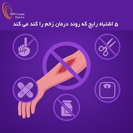
۰۲ اردیبهشت اخبار ۵ اشتباه رایج در مراقبت از زخمها که روند درمان را کند میکنداردیبهشت ۱۳, ۱۴۰۴ نویسنده مدیرسایت ۰ comments Facebook Twitter Pinterest linkedin WhatsApp Telegram ۵ اشتباه رایج در مراقبت از زخمها که روند درمان را کند میکند مقدمه زخمها از سادهترین خراشها تا زخمهای مزمن دیابتی، همگی نیاز...ادامه مطلب
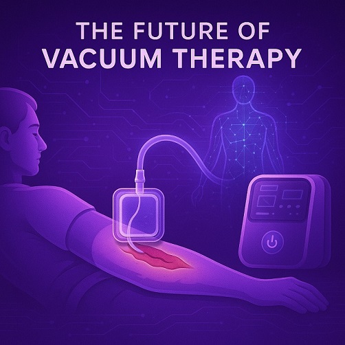
۲۵ فروردین اخبار, مقالات آینده روشن وکیوم تراپی؛ انقلابی در درمان زخمهای مزمناردیبهشت ۲, ۱۴۰۴ نویسنده مدیرسایت ۰ comments Facebook Twitter Pinterest linkedin WhatsApp Telegram مقدمه در دنیای امروز، با افزایش بیماریهای مزمن و مشکلاتی همچون دیابت، زخمهای فشاری و زخمهای جراحی، نیاز به روشهای درمانی نوین بی...ادامه مطلب
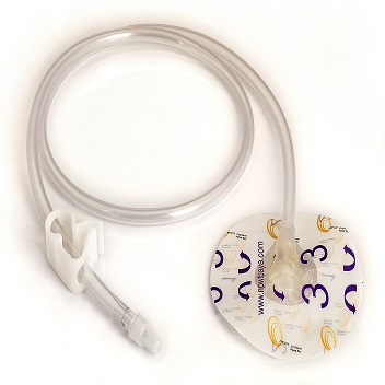
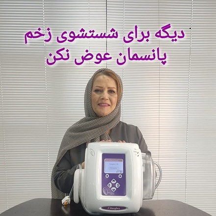
۲۱ آذر اخبار دیگه برای شستشوی زخم بیمارت پانسمان عوض نکن!آذر ۲۱, ۱۴۰۳ نویسنده مدیرسایت ۰ comments Facebook Twitter Pinterest linkedin WhatsApp Telegram JTNDZGl2JTIwaWQlM0QlMjIyNTE5Mzc1NjY0NiUyMiUzRSUzQ3NjcmlwdCUyMHR5cGUlM0QlMjJ0ZXh0JTJGSmF2YVNjcmlwdCUyMiUyMHNyYyUzRCUyMmh0dHBzJTNBJTJGJTJ...ادامه مطلب
۱۱ آذر اخبار آموزش روش کار با دستگاه وکیوم تراپی زخم Integro IIآذر ۱۱, ۱۴۰۳ نویسنده مدیرسایت ۰ comments Facebook Twitter Pinterest linkedin WhatsApp Telegram JTNDZGl2JTIwaWQlM0QlMjIyOTY4MDExMTQwMyUyMiUzRSUzQ3NjcmlwdCUyMHR5cGUlM0QlMjJ0ZXh0JTJGSmF2YVNjcmlwdCUyMiUyMHNyYyUzRCUyMmh0dHBzJTNBJTJGJTJ...ادامه مطلب